3D Mammography
Good Samaritan Medical Center Outpatient Imaging is excited to offer 3D Mammography
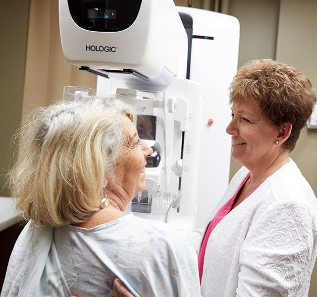
Good Samaritan Medical Center Outpatient Imaging is proud to offer advanced 3D mammography (breast tomosynthesis) designed for more accurate breast cancer screening. 3D mammography allows the radiologist to examine breast tissue one layer at a time, making fine details more visible because they are no longer hidden by tissue.
Increasing Cancer Detection with Less False Alarms
3D Mammography screening has shown an increase in cancer detection and a decrease in false alarms, according to a study published in The American Medical Association. Even more, the recall tests for women who had 3D mammography compared to women with conventional mammography was 36.6 percent lower, according to a study published by Yale School of Medicine. 3D mammograms will get you the answers you need, without the worry.
Benefits of 3D mammograms may include:
- Decrease in false alarms due to abnormalities
- Fine details are more visible and are less likely to be hidden by overlapping tissue
FAQs
Why is there a need for tomosynthesis breast exams? What are the benefits?
With conventional digital mammography, the radiologist is viewing all the complexities of your breast tissue in a flat image. Sometimes breast tissue can overlap, giving the illusion of normal breast tissue looking like an abnormal area.
By looking at the breast tissue in one millimeter slices, the radiologist can provide a more thorough assessment. In this way, 3D mammography finds cancers missed with conventional 2D mammography. It also means there is less chance your doctor will call you back later for a “second look,” because now they can see breast tissue more clearly.
What is a 3D mammography breast exam?
3D mammography is a revolutionary new screening and diagnostic tool designed for early breast cancer detection that can be done in conjunction with a traditional 2D digital mammogram.
During the 3D part of the exam, the X-ray arm sweeps in a slight arc over your breast, taking multiple breast images. Then, a computer produces a 3D image of your breast tissue in one millimeter slices, providing greater visibility for the radiologist to see breast detail. They can scroll through images of your entire breast like pages of a book.
The additional 3D images make it possible for a radiologist to gain a better understanding of your breast tissue during screening and may reduce the need for follow-up imaging.
What should I expect during the 3D mammography exam?
3D mammography complements standard 2D mammography and is performed at the same time with the same system. There is no additional compression required, and it only takes a few seconds longer for each view.
Is there more radiation dose?
Very low X-ray energy is used during the exam, just about the same amount as a traditional mammogram done on 2D.
Who can have a 3D mammography exam?
It is approved for all women who would be undergoing a standard mammogram, in both the screening and diagnostic settings.
What is the difference between a screening and diagnostic mammogram?
A screening mammogram is your annual mammogram that is done every year. Sometimes the radiologist may ask you to come back for follow-up images which is called a diagnostic mammogram to rule out an unclear area in the breast or if there is a breast complaint that needs to be evaluated.